Problem
Solution
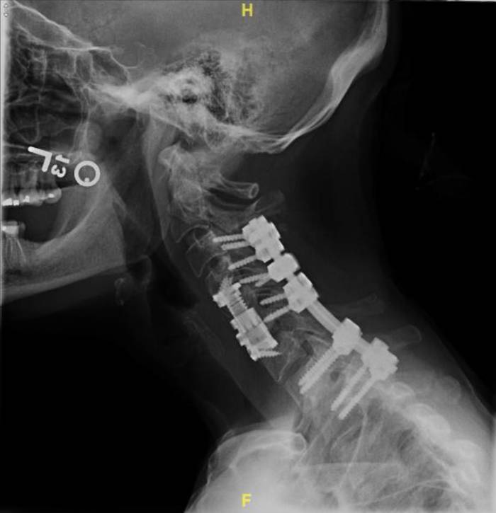
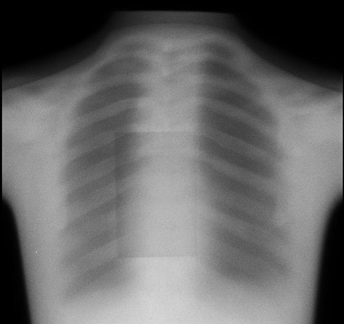
For both the lateral and the AP views, there are two separate sources of motion that could contribute to differences between the “pre” and “post” images: movement of the patient and the ovement of the spine and/or screws. The camera itself has preset positions for both views, with millimeter-level noise between separate images that can be effectively ignored. As the movements of the patient are extraneous, they introduce noise that makes it difficult to evaluate patient progress and diagnose any potential issues (e.g. pseudarthrosis) that may cause major health risks. As a result, a patient that may come in after their operation to diagnose why they haven’t seen the expected improvement may require additional x-rays which delay the process, which could result in any medical conditions worsening or potentially another operation being required.
We will create an algorithm enabling users to upload both the “pre” and “post” images, realign/manipulate them, and eliminate noise induced by movement of the patient. By doing so, the lab will be able to make more accurate diagnoses and prognoses of their patients. Having specific, quantitative data on the relative positions and movements of screws and vertebral bodies of interest would help patients to make the best of their own and the lab’s time and diagnose any complications earlier for better health and less cost to the patient.
Tech Stack


For all inquiries of partnership or sponsorship, please contact us at uiuc@hack4impact.org.